2 Types of Dental X-Rays

Dental x-rays are important diagnostic tools general dentists use to evaluate your underlying bone structure, plan for dental implants or extractions, and detect cavities. Part of your semi-annual dental checkup will most likely consist of having dental x-rays performed.
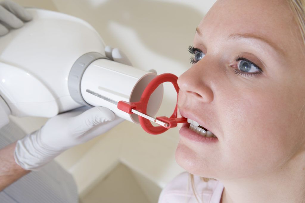
Dental x-rays don’t hurt, only take a few minutes to complete, and use extremely low amounts of radiation. When having a dental x-ray, you can expect to hold still and bite on certain plastic discs to obtain the proper angles to best view your teeth.
There are two main types of dental x-rays that may be performed: intraoral and extraoral. Intraoral x-rays focus on the inside of the mouth, while extraoral x-rays focus on the outside of the mouth. In most cases, intraoral x-rays are performed more often than extraoral x-rays. Within these two types of dental x-rays, there are also different types of intraoral and extraoral x-rays.
Types of Intraoral X-Rays

- Bite-Wing X-Rays can be used when placing a dental crown or filling, or to detect cavities between the teeth. Bite-Wing x-rays focus on the tooth’s crown from top to where it meets the bone. Details from the top and bottom teeth can be shown, but only in one particular area.
- Periapical X-Rays are used to diagnose issues in the root and surrounding bone structure. Periapical x-rays show the entire tooth from top to where the root meets the jawbone. All the teeth on the upper or lower arch can be shown in a single periapical x-ray.
- Occlusal X-rays show tooth placement of all the teeth in the upper or lower arch.
Types of Extraoral X-Rays

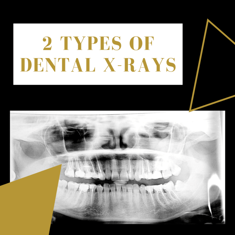
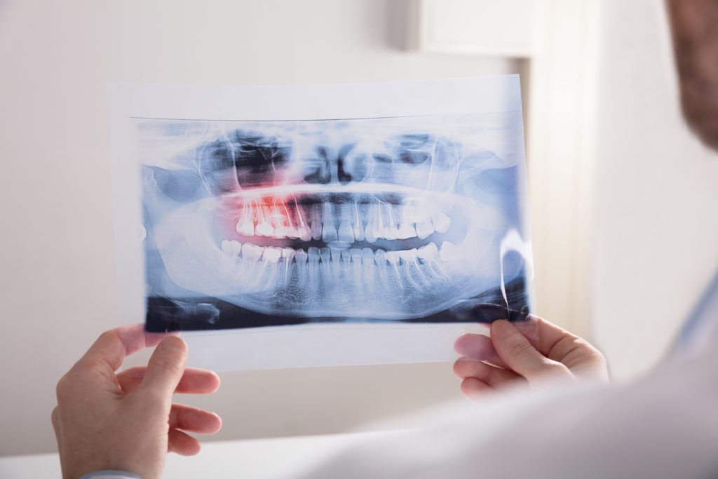
- Panoramic x-rays are used to view the position of teeth, detect emerging teeth, and identifying impacted wisdom teeth. Panoramic x-rays capture both the upper and lower arch in a single image.
- Tomograms are used to view structures that are hard to visualize because of their closeness to other structures. A tomogram isolates a single “slice” of the mouth and blurs all other layers.
- Cephalometric Projections show the side profile of the head to show how the teeth and jaw are aligned. These are commonly used prior to orthodontic treatment.
- Computed Tomography also known as a CBCT scan, is used in preparation for placing dental implants or performing tooth extractions. CBCT scans produce a single 3-dimensional image of the soft tissues, nerves, and bone of the mouth.
As you can see, there are several types of intraoral and extraoral x-rays that allow dentists to evaluate a number of dental issues. When visiting your dentist’s office for your semiannual dental checkup, you can most likely expect to have bite-wing, occlusal, panoramic, and perioaplical x-rays taken. More dental x-rays may be obtained once a treatment plan is decided upon.

Dr. John Batlle attended the UF College of Dentistry where he received his Doctor of Dental Medicine degree in 1983. After graduating, he worked for the State of Florida and received his commission in the Navy Reserve Dental Corps. He was deployed to Guantanamo Bay, Cuba in 2002 where he served as the dentist for Detainee Operations and Navy Hospital GTMO. He recently retired from the U.S. Navy Reserve after 26 years of service.

